Monday, 30 November 2009

John William Draper

John William Draper(1811-1822) was a scientist born in St Helens Merseyside and immigrated to USA . He became a chemistry professor in New York university. Most of his work was done in the field of photochemistry.He was one of the first scientists to use Daguerre's new invention of photography.
In March 1840, he presented the Lyceum of natural history in New York with the first representation of the moon's surface ever taken by photography in high quality by John William Draper.The silver platinum plate (Daguerreotype print) of the moon was,the first of
a series that where shot using a telescope. This series was later presented at the Science Academy.He was also the first to shoot portrait in America.
He made it the subject of special study, and was the first person in the world to apply it to individuals. "The first photographic portrait from life was made by me," he says, and "the face of the sitter," his sister Catherine
In 1872 he photographed the spectrum of a Lyrae (Vega), showing dark lines, a result then unique in science, and in 1873 the finest photograph of the diffraction spectrum ever made.

The First photo of the moon 1839


http://www.famousamericans.net/johnwilliamdraper/
http://www.fotoart.gr/photography/history/historyphotos/onephotoonestory/thefirstphotoofthemoon.htm

Pictures and diagrams

There are eight planets in our solar system :the so-called inner planets of Mercury,Venus,Earth,and Mars.Further away from the Sun are the gas giants Jupiter,Saturn Uranus and Neptune. The moon is the fifth largest satellite and also the only natural satellite in the Solar System.

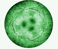
Most of the patients who I photograph after looking at the fundus photograph of eye mentioning the similarity of the mages with the Moon!Few weeks ago I saw an article in Guardian Newspaper about astronomy! I scan this photo from the paper.The more I look at the Imagery of Celestial objects, the better I can recognize their fascinating visual connection with medical fundus photographs .


Jupiter


Scars from the crash of Comet Shoemaker-Levy 9 appear on Jupiter's surface as a series of maroon blotches in this photo. The comet broke into 21 pieces before it hit Jupiter in 1994. Image credit: Hubble Space Telescope Comet Team and NASA


Ganymede, a moon of Jupiter, has craters and cracks on its surface. Asteroids and comets that hit Ganymede made the craters. The cracks are due to expansion and contraction of the surface. Image credit: NASA


The planet Jupiter's Great Red Spot is a huge mass of swirling gas. At its widest, it is about three times the diameter of the Earth. Image credit: NASA


The layers of dense clouds around Jupiter appear in a photograph of the planet taken by the Voyager 1 space probe. The large, oval-shaped mark on the clouds is the Great Red Spot. The spot is believed to be an intense atmospheric disturbance. Image credit: Jet Propulsion Laboratory


Callisto, a moon of Jupiter, is covered with craters produced when asteroids and comets struck its icy surface. Beneath the surface may be an ocean of salty liquid water. Image credit: NASA

http://www.nasa.gov/worldbook/jupiter_worldbook.html

our universe is not silence!


From an original CD: JUPITER NASA-VOYAGER SPACE SOUNDS (1990) BRAIN/MIND Research
Fascinating recording of Jupiter sounds (electromagnetic "voices") by NASA-Voyager. The complex interactions of charged electromagnetic particles from the solar wind , planetary magnetosphere etc. create vibration "soundscapes". It sounds very interesting, even scary.
Jupiter is mostly composed of hydrogen and helium. The entire planet is made of gas, with no solid surface under the atmosphere. The pressures and temperatures deep in Jupiter are so high that gases form a gradual transition into liquids which are gradually compressed into a metallic "plasma" in which the molecules have been stripped of their outer electrons. The winds of Jupiter are a thousand metres per second relative to the rotating interior. Jupiter's magnetic field is four thousand times stronger than Earth's, and is tipped by 11° degrees of axis spin. This causes the magnetic field to wobble, which has a profound effect on trapped electronically charged particles. This plasma of charged particles is accelerated beyond the magnetosphere of Jupiter to speeds of tens of thousands of kilometres per second. It is these magnetic particle vibrations which generate some of the sound you hear on this recording.
Sounds like a giant metal!
http://www.inner-net.com/bmr/wsounds/jupiter.wav
Each planet has its own classically composed theme.


Sunday, 22 November 2009

Ophthalmic photography history

Ophthalmic photography is a specific branch of scientific and medical photography that deals exclusively with eyes and related parts.this field in photography depends of anatomy and physiology of eyes as much as technique and technology in photography.
Modern imaging of the retina began in 1886 with Jackman and Webster who produced the first human retinal photographs in vivo. Many other contributions were made over the next 50+ years until two medical students (Novotny and Davis) at Indiana University
The first study of human Fluorescein radiograms happened in the late 1950s.


Dr. Harold Novotny a senior medical student began to think about passing some kind of dye through the system to determine saturation levels. It was thought that the observation of fluorescence might make it easier to make that determination, and sodium fluorescein was one of the dyes used.

Dr Novotny with his colleague Dr Alvis began performing fluorescein studies on diabetic patients, and individuals with hypertension. For the first time their work got rejected afterThey submitted their work to the American Journal of Ophthalmology in 1960, because of similar work with cinematography published by Dr. Milton Flocks and co-workers.They presented their technique for photographing fluorescence at the Association for Research in Ophthalmology on April 23, 1960, and after that an abstarct of their work was published in the American Journal of Ophthalmology in July, 1960. In July of 1961, the first published description of the technique appeared in Circulation.


http://www.opsweb.org/OpPhoto/Angio/FirstFA/FirstFA.html

Saturday, 21 November 2009

Mythology fo eye, representations in art


The Eye of Horus: http://www.ancientegyptonline.co.uk/eye.html

Creation stories:
http://www.historyworld.net/wrldhis/PlainTextHistories.asp?historyid=ab83

India: "In an early story Purusha is a primal man sacrificed by the gods as the act of creation. The sky comes from his head, the earth from his feet, the sun from his eye and the moon from his mind."

China: "After all this effort P'an Ku falls to pieces. His limbs become the mountains, his blood the rivers, his breath the wind and his voice the thunder. His two eyes are the sun and the moon. The parasites on his body are mankind."

Suns and Planets in Neolithic Rock Art:
http://www.maverickscience.com/arch-sun-planets.htm [Not sure this is useful, looks a bit 'whacky' to me]

Bosch, 'Saint John The Baptist on Patmos'.
http://en.wikipedia.org/wiki/St._John_the_Evangelist_on_Patmos

front of the image
back of the image


It looks to me like the reverse of the painting expands the colour and circular motif of the moon from the front and turns it into teh pupil of an eye. God's eye

Democritus,400BC,Ocular anatomy

Retinal imaging has evolved through many years. However, there are certain sentinel landmarks which have changed the way we view and understand the retina forever. The earliest of these occurred 400 years B.C. when Democritus first proposed the construction of the eye as a hollow tube connected to the brain .


Figure 1. Illustrations by Democritus (400 BC) on the left and Celsus on the right (from Duke-Elder, 1961).

Four hundred years later Celsus advanced the understanding of the ocular system to include a lens, the anterior chamber, and a vitreous .


http://www.ophthalmologyweb.com/FeaturedArticle.aspx?spid=23&aid=318

"Democntus explains sight by the visual image, which he describes in a peculiar way; the visual image does not arise directly in the pupil, but the air between the eye and the object of sight is contracted and stamped by the object seen and the seer; for from everything there is always a sort of effluence proceeding. So this air, which is solid and variously colored, appears in the eye, which is moist ; the eye does not admit the dense part, but the moist passes through." - http://www.aquinasonline.com/Magee/democritus.htm


"Vision works by the eye receiving "images" or "effluences" that are emanated by bodies." - http://www.newworldencyclopedia.org/entry/Democritus


The History of medical illustration

The Egyptians pictured their civilisation on papyrus rolls and sculptured reliefs and produced the world's first surgical textbook around the time of Tutankhamen-1700BC.It describes in detail 48 cases of battle wounds and their treatments.
the Greek artists,like the Egyptians used everyday themes in their decorative arts and most aspects of their culture and medicine can be found in painting on their black figure pottery.dating from 400BC
After the fall of Roman Empire civilisation slipped into the Dark Ages. Christaniaty emphasis on the soul and not the body,medical investigation virtually stopped.
Until the 14th century medicine was taught exclusively on the text of Galen who had died 1100 years before. because his work was approved by church.
Universities were growing fast in the 14th and 15th century.Leonardo de vinci's approach to anatomy was the beginning of modern scientific method,with controlled investigation,detailed recording and accurate illustration of finding.( more explanation in one of my earlier post)


Illustration has been an important feature of medical documentation since the time of Vesalius and thus has a long history. However, the first application of photography to medicine appears in 1840, when Alfred Donné of Paris photographed sections of bones, teeth, and red blood cells using an instrument called the microscope-daguerreotype. Conventional medical photography apparently began in France when J. G. F. Baillarger photographed cretins (1851), which was followed by a Dr. Behrendt of Berlin photographing his orthopedic cases in 1852, and in the same year by Dr. Hugh Welch Diamond photographing mental patients at the Surrey County Asylum in England.

When E. J. Muybridge synthesized motion studies (chronophotog-raphy) of humans and animals (1877–1893) a new area in medical photography occurred, which greatly stimulated other investigators in medical photography, and the French physician Étienne-Jules Marey endeavored to analyze human and animal motion by serial photographic studies (1882) and devised a chronophotographic apparatus and projector (1890), which was the forerunner of the modern motion-picture camera. By the turn of the century, applications were too numerous to mention, but in 1927 R. P. Loveland made a medical teaching film using cinephot-omicrography, demonstrating the life history of the yellow fever mosquito, and in 1929 F. Neumann in Germany made a time-lapse film of living bacteria.

The first medical publications illustrated with photographs were Album de Photographies Pathologiques and Mecanisme de la Physiologie Humaine (1862) by G. B. Duchenne, the founder of electrotherapy. The Photographic Review of Medicine and Surgery (1870), by F. F. Muary and L.A. Duhring, was the first medical journal illustrated with photographs. Albert Londe in 1888 published a book, La Photographie Moderne , containing information on medical photography and also the first book specifically devoted to medical photography, La Photographie Medicale in 1893.


Journal of Audiovisual media in medicine 1986,9,44-49 printed in Great Britain

http://encyclopedia.jrank.org/articles/pages/1156/Medical-Photography.html

Thursday, 12 November 2009

Dominant ideas

I’ve been working as an Ophthalmic Photographer for 2 months at St Helens Hospital. I photograph eyes.

One of the less obvious, but extremely important, common features of eyes is the way they move, either by themselves or as part of a moveable head or body

My interest in science lead me to develop and combine my two loves, looking at the narratives with the technical expertise. Lennart Nilsson who is regarded, as the greatest scientific photographer in the world has been my main inspiration in scientific photography

Before starting this job I never thought how much we take our visual abilities for granted. The more I learn about eyes the more I realize how fascinating, amazing,suite of abilities and mysteries it is.These images are Powerful and emotive. I am hoping to visualize my appreciation about this beautiful part of body to the others who aren’t able to see with them with naked eyes!

In my previous posts Re: Topcon TRC, Florescent Digital Imaging I explained about Fundus imaging and florescent angiograms, which are the images I am concentrating on. I am hoping to visualise the diversity and beauty existed in scientific/ fundus images by messing with them artistically by using my photographic talent mixed with media and animation.

I have recognised a fascinating visual connection between medical fundus photographs (of the back of the eye) and imagery of Celestial objects such as Jupiter. I intend for my work to reflect such visual similarities and hope to be able to capture the sense of wonder I feel towards astronomical objects and the intricacies of the human body.

Few ideas how to present it :


PJSaine/an ophthalmic photographer

During my reaserch i came acorss PJSaine who is an Ophtalmic photographer for 20 years.
"Ophthalmic photography allows me to use my photographic talents to help people by gathering visual information for specialists in ophthalmology .Mostly I photograph retinas, but I also image corneas and lenses, the cells lining the inside of the cornea, and the facial area around eyes. I perform dye tests such as fluorescein angiography (FA) and ICG angiography (ICG-A), as well as specialized laser based testing such as Ocular Coherence Tomography (OCT)."

he mentioned W.Eugene Smith as his main inspiration.W.Eugene Smith wanted his photographs of WWII to be an indictment of war. He wanted his images to change human behavior by showing people the horrors of armed conflict. Our nation's more recent history - from Korea to Vietnam to Iraq - demonstrates that he did not (possibly could not?) accomplish his goal. "I chose a smaller, but still worthwhile goal: to use my photography skills as part of a team that helps people see better"said PJSaine.The other reason he chose Ophtalmic photography as a carrier was a newness of the proffesion.

Here are some examples of his images that i found quite intresting the way he combines his artistic expertise with his photographic and scientific technique.


Celtic Quilt TriColor Retina


Retina rings


http://www.pjsaine.com/OphthalmicPhotography/PJSOphth.html

Thursday, 5 November 2009

Software!

http://videomaker.com/community/blogs/freeware/jahshaka-freeware-a-list/

http://jahshaka.org/

http://www.stereoeye.jp/software/index_e.html

recently I started playing around with a new software called After effect which is a digital motion graphics and compositing software.
I started reading the tutorial about this software.I am not expecting to be a master for this programme at the end of this module but I am hoping with a help from one of the graphic design student to create a motion graphics and visual effects form the fundus photograph of eye in comparison with planet ideally Jupiter because of the visual similarity.